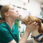
The dog’s eyes are a complex apparatus that makes it possible for them to see the beautiful world we live in. Unfortunately, they are also very sensitive organs and can be affected by many different conditions.
In This Article You Will Read About
Sometimes the problems come from the eyes themselves and sometimes the eye problem is secondary to some other disease. What to do when your pet has a dog eye disease?
Dog eye disease should always be taken seriously. When you think your dog has such a condition you need to react fast and prevent loss of vision. A trip to the veterinarian’s office is imminent!
This overview of the most commons eye problems in dogs will give you a general idea of what might happen and how serious it can get.
The most common eye conditions in dogs are:
- Cherry Eye
- Corneal Wounds
- Pink Eye (Conjunctivitis)
- Dry Eye (Keratoconjunctivitis Sicca – KCS)
- Cataracts
- Glaucoma
- Progressive Retinal Atrophy (PRA)
Cherry Eye
Dogs and many other mammals have an extra eyelid called a third eyelid or nictitating membrane. It’s located in the inner corner of the eye within the lower eyelid.
The nictitating membrane has the role of an additional layer for the eye that protects it from injuries during fighting or hunting. A special gland inside this eyelid produces a tear film for the eye.
Sometimes the gland prolapses or pops out of place. When this happens the condition is called cherry eye. It can happen only on one eye or bilaterally.
The gland is anchored by fibrous tissue to the inner lower rim of the eye. In some breeds like Shih Tzus, Lhasa Apsos, Bloodhounds, Boston Terriers, Beagles, Cocker Spaniels, and Bulldogs this fibrous attachment is weak and allows the prolapse.
Symptoms, diagnosis, and treatment of Cherry Eye
If your dog has a red and swollen mass in the corner of the eye it probably has a prolapsed third eyelid gland. The mass can be big and inflamed and cover a large part of the cornea or be small and insignificant.
Even though it’s not considered to be an emergency you should consult your veterinarian as soon as possible.
The treatment protocol is to reposition the gland within the third eyelid. It’s advisable to decide on surgery sooner because untreated Cheery Eye can cause permanent eye damage. Almost 50% of the liquid portion of the tear film is released from this gland.
Without its function, the eye will sooner or later dry out. Within weeks of surgery, the gland returns to its normal function. In 20% of surgically treated cases of a cherry eye the gland prolapses again.
With chronic and severe cases the only option left might be to completely remove the gland, especially if its function is already diminished.
Corneal Wounds
The shiny and transparent membrane on the front of the eyeball is called a cornea. It’s consisted of three very thin layers of cells aligned one over the other. The outermost layer is the epithelium; beneath it is the stroma with the Descemet’s membrane at the bottom.
Corneal wounds are a result of physical injury. When only a few epithelium layers are damaged the condition is called corneal abrasion or corneal erosion. Deeper forms when the damage goes throughout all of the layers are referred to as corneal ulcers.
Damage of the stroma and the Descemet’s membrane is called descemetocele. This is a very serious injury because a ruptured membrane lets all the liquid in the eyeball leak out.
Trauma is the most common cause of corneal wounds. Sharp trauma (cat scratch, sharp object) and blunt trauma (rubbing the eyes) can both be responsible for the damage.
Different chemicals and shampoos or foreign bodies are causative agents as well. Primary or secondary bacterial and viral infections of the eye are less common causes of corneal defects.
Symptoms, diagnosis, and treatment of Corneal Wounds
Corneal wounds are very painful and most dogs in an attempt to relieve the pain will continuously rub the affected eye. They keep their eyelids tightly closed to protect the eye and on occasions, discharge can be seen.
Veterinarians use special dying techniques to determine the depth of the wound. Tiny defects can only be seen with ophthalmic light and filters that enhance the point of view.
Abrasions and ulcers are usually treated with eye drops and ointments that help with infection control and let the eye heal on its own. Dogs can’t wear eye patches to sometimes surgically stitching the upper and lower eyelid is needed until the eye gets better.
Pink Eye (Conjunctivitis)
Conjunctivitis is inflammation of the mucous membrane that covers the inner eyelids, the eyeball, and the third eyelid. This membrane is similar in structure and function to the lining of the mouth and the nose.
Pink eye it’s most commonly a result of viral infections (distemper virus), but can also be due to allergies, autoimmune disorders, tumors, poor tear production, obstructed tear ducts, trauma, foreign bodies, etc.
Collies and Collie-breed mixed dogs have a condition called nodular episcleritis that can lead to pink eye. Plasma cell conjunctivitis in German Shepherds is another breed-related cause.
Symptoms, diagnosis, and treatment of Pink Eye
The most prevalent symptom is eye discharge which can be yellow, green, or cloudy. The areas around the eyes are swelled and red and the dogs are blinking or squinting excessively.
Dogs can have conjunctivitis on one eye or both eyes. Other clinical signs associated with conjunctivitis are coughing, nasal discharge, and sneezing.
The main goal of every veterinarian is to determine if conjunctivitis is a primary or a secondary problem. To give a precise diagnosis the dog needs to go through a detailed ophthalmic examination.
Treatment is pointed towards the actual cause of Pink Eye. In primary conjunctivitis, the dogs will be treated with topical or oral medications like antibiotics, NSAID’s or corticosteroids.
Most dogs will recover from the disease perfectly. Chronic problems and some immune-mediated cases of Pink Eye require life-long therapy.
Dry Eye (Keratoconjunctivitis Sicca – KCS)
When there isn’t enough tear film to keep the eye wet the cornea and surrounding tissues become inflamed. The tear film consists of water, fatty liquid, and mucus.
Without enough film to lubricate the structures on the eye infective microorganisms and debris can contract the eye.
Any condition that interferes with tear film production will eventually contribute to the development of the dry eye.
Sometimes the dog’s immune system attacks some of the cells that produce a fraction of the tear film. Other causes are inner ear infection (with nervous system effects), hypothyroidism, sulphonamides (medications), viral infections, etc.
Breeds predisposed to Keratoconjunctivitis Sicca are:
- Samoyed
- Pug
- Pekingese
- English Bulldog
- Boston Terrier
- Lhasa Apso
- English Springer Spaniel
- Cavalier King Charles Spaniel
- Miniature Schnauzer
- Shih Tzu
- Yorkshire Terrier
Symptoms, diagnosis, and treatment of Dry Eye
Dogs KCS have irritated red and painful eyes. Sometimes they hold their eyes constantly shut and other times they blink excessively.
When the watery part of the film is missing you can spot thick and yellow discharge on the eyes. Often there are ulcers on the cornea. Corneal damage is the reason for hyperpigmentation and black scarring.
Based on the clinical signs and medical history your vet will decide whether or not to perform the tear production tests called Schirmer’s tear test. Special wicking paper is used to measure how much tear film is produced in a minute.
The dry eye requires treatment for a lifetime. The two main goals are to replace tear film and to stimulate the production of tears. New generation eye medications like tacrolimus and cyclosporine are easily applicable and very effective in treating the condition.
Antibiotics and pain-relief medications can also be used when there are secondary issues. The prognosis for dry eye is great when you carefully follow the vet’s instructions.
Cataracts
Cataracts are a cloudy film that forms on the lenses of the eyes and prevents light from entering. They are a product of the clumping of eye proteins.
Cataracts usually start as small protein accumulations and grow more and more. There are cases though when they appear overnight and blind the dog completely.
There is an inherited trait of cataracts in dogs. Diabetes mellitus, eye injuries, and inflammations of all sorts can contribute as well. Usually, this is a condition most prevalent in senior dogs.
According to a study the prevalence of Cataracts is 26% in dogs aged 9 to 11.
Symptoms, diagnosis, and treatment of Cataracts
Symptoms of cataracts in dogs include:
- Changes in eye color
- Changes in pupil shape or size
- Cloudy pupils (one of both eyes)
- Difficulty seeing in low light
- Clumsy behavior
- Scratching/rubbing of the eyes
Veterinarians use a bright light and magnifying lenses to detect the formation of cataracts. They can be seen when they are still immature and haven’t started to impair the dog’s vision just yet. That’s why regular check-ups are important, especially in older dogs.
The only therapeutic solution is the surgical removal of cataracts. Although they are not fatal and most dogs learn to live with the symptoms, it’s better to go for surgery and prevent blindness – it’s for the sake of the dog’s quality of life.
Glaucoma
Glaucoma is an eye condition when the pressure inside the eye (intraocular pressure) is increased. When the production and drainage of aqueous humor inside the eye are disrupted, the pressure builds up.
Primary glaucoma happens in certain dog breed due to inherited anatomical abnormalities which make the drainage of fluid hard. Secondary glaucoma is a result of eye injury or some other systemic disease.
Common causes of glaucoma in dogs are bleeding inside the eye, damaged lenses, dislocated lenses, tumors, uveitis (eye inflammation), etc. When the pressure is permanently increased degenerative changes to the optic nerve and the retina happen.
Symptoms, diagnosis, and treatment of Glaucoma
Common signs of glaucoma in dogs are:
- Pain in the eye
- Discharge (watery)
- Loss of appetite
- Lethargy
- Physical swelling around the eye
- Cloudy or bluish eye
- Blindness
All of the signs can develop pretty quickly in acute cases.
Diagnosis is made by measuring the intraocular pressure with a special ophthalmic instrument called a tonometer. All cases of acute glaucoma are an emergency.
The key goal of the treatment is to decrease the pressure as soon as possible to eliminate the risk of severe damage to the eye. If there is an underlying disease responsible for glaucoma it needs to be treated at the same time.
Veterinarians prescribe drugs that promote drainage and meds for pain control. In advanced cases, surgery can be the only possible solution. Removal of the eye is recommended when the dog is already blind.
Progressive Retinal Atrophy
The retina is a group of cells at the back of the dog’s eye that’s responsible for converting light signals. With progressive retinal atrophy the retina wastes over time, the photoreceptor cells degenerate and the dog ends up blind.
PRA is an inherited disease that the dog must get from both parents. Commonly affected dog breeds are:
- Rottweilers
- Bullmastiffs
- Old English Mastiffs
- Golden Retrievers
- Labradors
- Cavalier King Charles Spaniel
Dogs that develop PRA shouldn’t be used for breeding. The onset of symptoms is noted in dogs 3-9 years of age. There is another form of the disease called retinal dysplasia which appears in puppies 12-16 weeks of age.
Symptoms, diagnosis, and treatment of PRA
The condition is not painful at all. The first symptom is night blindness and affected pups have problems while moving in dark rooms. Their pupils are dilated more than usual during the night and light shines on them.
As the disease progresses the dogs become clumsy during the daytime as well and eventually completely lose their vision. It takes about 1-2 years for the dog to become blind.
Veterinary ophthalmologists use sophisticated testing called ERG (electroretinogram) that can diagnose PRA even before the onset of the symptoms.
Sadly there is no effective treatment for this condition. Vitamins and antioxidant supplements can delay the outcome but won’t prevent blindness.
Conclusion
Eye diseases in dogs are very common and can vary from mild to very serious. Some dogs continuously have them throughout their whole lives, but still manage to live normally. The eye examination should be a part of the annual checkup plan.
Rottie owner? Get your Rottie featured on our site with a dedicated page…
Simply fill out the form in the link with a picture and description and we’ll create a dedicated page on our site featuring your Rottie.

